Oral Pathology Digital Radiography
This section will provide you with a short description of the different types of imaging instrumentation that our office provides. Over the years our offices have always been equipped with the most up to date imaging equipment available to provide our patients with the most efficient, dependable images with the least amount of radiation exposure. Today the imaging studies available have evolved dramatically as the type and quality of the images that are available is quite impressive. We have maintained our “state of the art” reputation and recently upgraded all units throughout our practice. In addition our offices now, are all totally linked electronically, so all radiographic studies performed at any location are readily available at the time of your office visit.
Currently our offices are 75% digitalized and we are equipped to perform the following film studies within the confines of our offices:
Periapical radiograph: This x-ray is the conventional dental radiograph (small dental radiographs) and is used in our office on occasion.
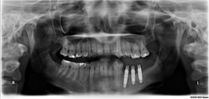
Orthopantomogram (Panorex): Panoramic imaging remains the standard of care for many types of dental procedures. In todays medical world the panorex is without question the most comprehensive preliminary screening imaging study available. It is essential for almost any type of oral surgical procedure. This study provides us with a complete over view of the lower 1/3 of the facial skeleton provides an enormous amount of diagnostic information with minimum radiation exposure. It remains the back bone of oral surgery and the least invasive (with respect to radiation exposure and patient acceptance). Panoramic units are in place at all three office locations and are of the highest quality, emitting the least amount of radiation while providing us with the necessary diagnostic information.
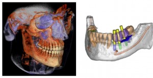
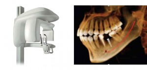
Our most recent advancement is our Cone Beam Computerized Tomographic (CBCT) machine located in the Hackensack office. This unit is a Kodak 9300CT and can provide a fully digitized 3-D study of the entire facial skeleton. The Kodak 9300CT is the most sophisticated cone beam unit available on todays market and while these units are becoming popular, there are a number of distinguishing features about our unit which make it the most sophisticated unit available at this time:
- The lowest level of radiation emissions per study.
- The 9300CT enables us to isolate our study, if necessary, to a small area of interest rather than “shoot the whole face”. From our point of view this feature is critical with respect to our patients overall health and welfare as we are able to effectively minimize radiation exposure to our patients and scan small isolated areas throughout the facial skeleton when necessary.
This new addition to our diagnostic armamentarium provides us with cutting edge technology that enables us to provide a clear and more definitive diagnosis (especially with respect to implant restoration and facial reconstruction) and more accurately anticipate potential surgical sequel.